(a) Prosoma of a theridiid spider, longitudinal section. The large venom glands (VG) extend far into the carapace. Chel = chelicera, Clyp = clypeus, SEG = supraesophageal ganglion. 100x. (b) A strong muscle layer (M) surrounds the body of the venom gland. 200x. (c) The wall of the venom gland in longitudinal section: the muscle layer (M) is connected via a basal membrane (BM) to the glandular cells (G). Secretory droplets (S) containing venom accumulate toward the lumen of the gland. 2,800 x.
(A)一种球蛛的前体,纵切面。大毒腺(VG)延伸到头胸甲内很远。Chel=chelicera,CLYP=clypeus,SEG=食管上神经节。100倍。
(B)强烈的肌肉层(M)包围着毒腺的本体。200倍。
(C)毒腺纵切面壁:肌层(M)通过基膜(BM)与腺细胞(G)相连。含有毒液的分泌液滴(S)向着腺体的管腔聚集。2800 x。
In the large tarantulas (mygalomorphs), the venom glands are quite small and lie inside the chelicerae (fig. 3.3a). In contrast, most labidognath spiders have relatively large venom glands that may extend out of the chelicerae and reach far into the cephalothorax (fig. 3.4a). In some extreme cases (Filistata, for example), the glands may be even larger and subdivided into lobules. The most curious specialization is found in the spitting spider Scytodes (fig. 3.5), in which the gland consists of an anterior part that produces the venom and a posterior part that produces silk and a gluelike substance (Kovoor and Zylberberg, 1972). Scytodes catches its prey by quickly spitting silk and glue onto it (Monterosso, 1928; Dabelow, 1958), thereby both entangling the prey’s appendages and fixing it to the ground. High speed video images (1000 frames/s) showed that the silk-glue ejection is very fast, close to 30 m/s, and lasts only for 30 ms (Suter and Stratton, 2009). Upon contact with the prey, the silk contracts by about 50%, which helps immobilize the prey. The slow- motion pictures also revealed how the zigzag pattern (fig. 3.6) comes about: the chelicerae are lifted upward, producing the vertical component of the pattern, while a rapid oscillation (300–1800 Hz) of the cheliceral fangs produces the horizontal spread. It is still unclear whether the ejected venom has any effect on the captured prey (Clements and Li, 2005).
在大型捕鸟蛛(原蛛下目)中,毒腺相当小,位于螯肢内(fig.3.3a)。相反,大多数labidognath蜘蛛都有相对较大的毒腺,这些毒腺可以延伸出螯肢并延伸到很远的头胸(fig.3.4a)。在某些极端情况下(例如,Filistata),腺体可能更大,并细分成小叶。最奇特的特化是在吐唾液的蜘蛛Scydes(fig.3.5)中发现的,它的腺体由产生毒液的前部和产生丝和胶状物质的后部组成(Kovoor and Zylberberg, 1972)。Scydes通过快速吐出丝和胶水来捕捉猎物(Monterosso, 1928; Dabelow, 1958),从而既缠绕了猎物的附肢,又将猎物固定到地面。高速视频图像(1000帧/秒)显示,丝胶喷射非常快,接近30m/s,持续时间仅为30ms(Suter和Stratton,2009)。当与猎物接触时,丝收缩约50%,这有助于固定猎物。慢动作图片还揭示了之字形图案(fig.3.6)是如何产生的:螯肢向上抬起,产生图案的垂直分量,而螯牙的快速摆动(300-1800 Hz)产生水平扩展。目前还不清楚喷出的毒液是否对捕获的猎物有任何影响(Clements and Li,2005)。
Chemically, spider venom is heterogeneous in that it may contain many different substances. It is a mixture mostly of large, neurotoxic polypeptides (molecular weight 5,000–13,000) and smaller biogenic amines and amino acids; proteolytic enzymes may also be present (Kaiser and Raab, 1967; Habermehl, 1975; Bachmann, 1976). Because almost all spiders have venom glands, they are all potentially poisonous—at least with regard to their normal prey. However, only about 200 of the 40,000 species of spiders are dangerously poisonous to humans (Maretic, 1975; Diaz, 2004). And only four genera (Atrax, Latrodectus, Loxosceles, Phoneutria) are known to cause potentially deadly bites (Isbister et al., 2003).
在化学上,蜘蛛毒液是异质的,因为它可能含有许多不同的物质。它主要是大的神经毒性多肽(分子量5,000-13,000)和较小的生物胺和氨基酸的混合物;也可能存在蛋白水解酶(Kaiser和Raab,1967;Habermehl,1975;Bachmann,1976)。因为几乎所有的蜘蛛都有毒腺,所以它们都有潜在的毒性--至少就它们正常的猎物而言是这样。然而,在40,000种蜘蛛中,只有大约200种对人类有危险(Maretic,1975;Diaz,2004)。只有四个属(Atrax,Latrodectus,Loxosceles,Phoneutria)已知会引起潜在的致命咬伤(Isbister et al., 2003)。
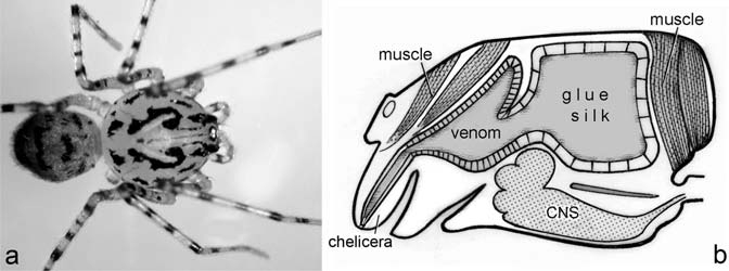
Figure 3.5


推荐文章


上一篇:科学网期刊编辑该让作者推荐审稿人吗? 下一篇:科学网Secondary aperture模型开发助力制备高性能G